How Is Gout Diagnosed?

Early diagnosis and treatment of gout are essential to avoid complications. If suitable medication and lifestyle changes are not adopted in time, the symptoms may become chronic and severe. Diagnosis of gout is difficult because similar symptoms appear in other forms of arthritis. A careful medical and physical examination is, therefore, a necessity.
Gout is typically diagnosed based on the symptoms experienced by the individual, plus their medical history and that of their family. These facts will be able to inform the doctor of the possibility of a case of gout but to rule out other causes of the symptoms, X-rays, and other scans may be used to eliminate other causes.

Signs and Symptoms of Gout
Pain and swelling in the big toe
Most people will experience their first gout attack in their big toe. It will usually be an excruciating pain radiating from the joint of the big toe that can be so bad it becomes debilitating. There will also be a swelling from the same joint that is red, tender and warm. This is usually a symptom that can easily be associated with gout, though gout can affect any other joint in the body.
One of the distinguishing features of gout is that after the initial attack, the symptoms may subside automatically within 10-12 days. If left untreated, a subsequent attack may occur after 5-6 months. In almost ninety percent of the patients, initially, it affects the lower extremities.
Pseudogout
This happens when gout affects other joints in the body that aren’t in the big toe. However, these will often be in the lower body, particularly the knee, but also the wrist, elbows, and ankles. Regardless of the location, the symptoms will always be the same: severe pain radiating from the joint; followed by swelling around the joint that is red and warm.
Key differences between Gout and Pseudogout:
In most of the patients, gout affects the big toe initially. Heel, fingertips, and wrists may be affected in some of the cases. On the other hand, Pseudogout almost always hits the knee first. Although preliminary infection may develop in the ankle, shoulder, wrist or the hip in a few instances.
Gout is caused by the deposition of abnormal levels of uric acid. Whereas, pseudogout is mainly caused by high levels of calcium pyrophosphate.
Gout is more prevalent in the age groups of 30 to 50. It is two times more common in men. Gender and ethnicity determine the level of risk for gout. While, Pseudogout affects people above the age of 60. Gender or racial background have no known correlation to pseudogout.
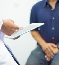
Tests for Gout
Physical Examination
The doctor carries out a complete physical examination of the patient to establish the exact nature of symptoms. The medical and family history of the patient is also considered to include all plausible causes. This examination is usually followed by a blood test to check the level of uric acid in the body.
Hyperuricemia
This is a condition where there is an unusually high level of uric acid in your body. The normal range for uric acid in the blood plasma is around 3.4-7.2mg/dL in men and 2.4-6.1mg/dL in women. The level of uric acid in the body can be determined through blood and urine tests that compare your levels to those of a similar healthy individual.
Although high uric acid levels may be indicative of gout, they don’t automatically mean that you have gout. Some people may have hyperuricemia or hypouricemia and not develop any gout symptoms, so it may be necessary to consider other symptoms.
Joint Fluid Analysis
This is a test for the analysis of the synovial fluid around the joints. The object of the test is to determine the presence of uric acid crystals in the joint fluid. This is the only clear method to diagnose for gout and eliminate all other forms of arthritis.
A long, thin needle is inserted into the joint. A syringe attached to the needle is used to extract synovial fluid. The process is carried out under a local anesthetic to eliminate pain. However, if you are not given anesthesia you may feel pain and discomfort. The process of extraction of synovial fluid is called Arthrocentesis. This fluid is sent to the lab for analysis.
A tight pressure bandage will then be applied to the joint to prevent swelling or bruising. After the procedure, you should rub ice over the area to relieve the soreness. A cortisone shot may be given to keep the fluid from building up again.
The procedure does not involve any major risks. You may experience a slight stiffness of the joint which subsides over time. In a few cases, there may be bleeding or infection that requires medical attention. Overall, complications are rare.
X- Ray Imagery
X-rays are sometimes used to diagnose for gout, but they aren’t very effective. In the early stages of gout, there is usually not a lot of damage to the cartilage or bone that can be seen by an X-ray. It may, therefore, not be very informative. However, in the late stages, the X-rays will give a clear diagnosis because the damage to the bone will be visible.
Advanced imaging technology have given a whole new direction to early diagnosis and treatment of gout. Although it is still in its nascent stage and there are no confirmed studies to prove its efficacy over the traditional methods of diagnosis.
Ultrasounds and Magnetic Resonance Imaging have grown in popularity in the recent years as diagnostic procedures. Ultrasounds are proving to be quite an effective tool. They can detect small crystals that may have been hiding under ligaments.
Advanced imaging can assist in the investigation of joint pathology in gout. It is also useful for early diagnosis of gout where x-ray imaging has failed. These techniques are being increasingly applied to monitor joint inflammation and damage during the course of treatment. There is a drawback though that these techniques are expensive and are not available easily.
A combination of traditional diagnostic methods along with MRIs is currently being employed to deliver optimum benefits to the patients.
The current benchmark in diagnosis is the presence of monosodium urate crystals in the joint fluid or tophus.