What Is Nodular Melanoma?
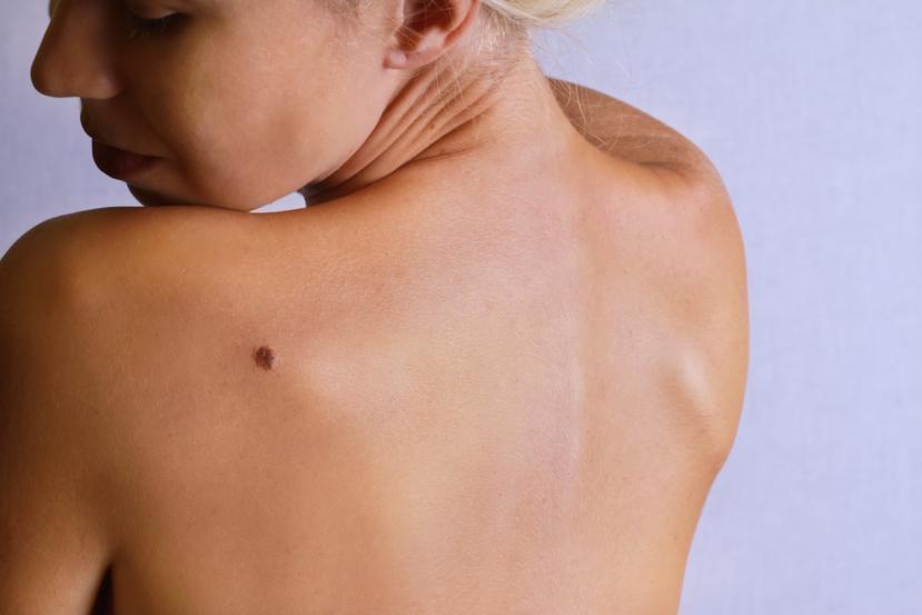
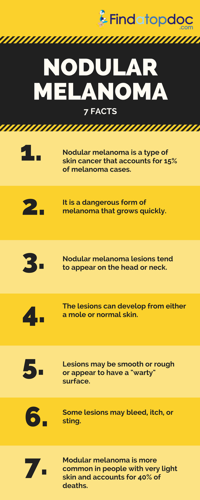
Melanoma is a serious skin cancer which arises from pigment cells called as melanocytes. An invasive form of melanoma is called nodular melanoma. Nodular melanoma is a serious skin cancer that got its name from the word "nodule", a doctor’s term for a lump as nodular melanoma lesions are shaped like a lump. Nodular melanoma lesions tend to appear on the head or neck. They can develop from an existing mole or grow out on normal skin.
Nodular melanoma is commonly characterized by cells that grow downwards into the skin in a vertical fashion. This type of skin lesion generally shows up as a type of nodule or lump that penetrates deeply into the skin and will show as a lump above the skin. Most nodular melanoma lesions are circular or roughly oval in shape. It is often larger than most moles and may be black, brown, red, or have varied colors. The lesion may be either smooth or rough or have crusty or warty surface. In some cases, an open wound (ulceration) appears above the lesion, or it may bleed. Some patients find that the lesion may itch or sting.


Nodular melanoma is more dangerous than ordinary melanoma. It grows rapidly and readily invades nearby tissues. The lesion grows by "burrowing" deep into the skin, and spreading (metastasis) is more likely to happen once it gains access to a lymph node. A nodular melanoma lesion grows noticeably in diameter and thickness within months. Nodular melanoma is very rare, accounting only for 15% of melanoma cases. Because it is aggressive, it affects more people and accounts for 40 percent of deaths. It is thought that changes in the DNA may be responsible for the speed of growth of these nodules.
It is found to be more common in people with very light skin and those who tan quite easily and occasionally in people with brown or black skin.
Nodular melanoma is the second most common subtype of melanoma and it accounts for 15 to 30 percent of all melanomas. Unlike the other melanoma subtypes, nodular melanoma appears to lack an initial rapid growth rate instead it begins with vertical growth. Nodular melanoma is more commonly seen in older people whose age above 50 years.
The lesions are mostly found in the head and neck. It arises in normal skin, but without intervening radial growth phase. It hardly takes three months for these type of cancers to spread internally.
Nodular melanoma can quickly move to the advanced stage, hence these type of skin cancers are very deadly. If the nodular melanoma is detected in the early stages, the doctors can look for some treatment, however in the advanced stages, nodular melanoma gets difficult to be successfully treated.
What are the Symptoms of Nodular Melanoma?
- Nodular melanomas are likely to be asymmetrical when you compare it to normal moles.
- Nodular melanomas would be found on the neck, head and trunk of the body. Nodular melanomas do not develop from an existing mole, it typically begins as a new growth altogether and then multiplies in number.
- Nodular melanoma would have fuzzy borders and notched or scalloped edges.
- At times, nodular melanoma would be just one colour. But most of the nodular melanomas shall appear as blackish-blue or reddish blue. Nodules which are flesh toned are called amelanotic nodules. Since the nodules lack pigment, the melanoma spots appear the same colour as the surrounding skin.
- If the skin lesion is larger than 6 mm in diameter or is growing, then it’s a sign of melanoma.
- Skin cancers begin as bumps or thick spots on the skin. The primary characteristic of nodular melanoma is that it’s a dome shaped growth on the skin just as the name suggests.
- Any birthmarks or moles on the skin which rise above the skin are typically limp or give easily when pressed. However nodular melanomas are not. Moles of nodular melanomas are firm when touched. They do not move when pressed with a finger. If you come across any such growth on your skin, press it with your finger, if you feel hard or a knot on that area then ask your doctor to check the growth.
- The growth of nodular melanomas is very quick. Normal moles initially develop and then stop growing within a few weeks. However, this is not the case with melanoma. New developments that continue to grow even after two to three weeks can be sign of a melanoma.
What are the risk factors for nodular melanoma?
Anyone who has skin that burns easily is more likely to get nodular melanoma. Moreover, it is also seen more often in men and in people who have already had another type of melanoma. If you are a person who has numerous moles, then there is a chance for nodular melanoma to grow in your body.
People with the following factors may also face an increased risk of having nodular melanoma:
- Previous history of having melanoma
- Having many moles
- Having one or more moles that do not look like normal moles in the body (atypical moles)
- Fair skin
- People who are 35 years old and older (although cancer can still occur in younger individuals)
Prognosis
The patient must always look for any new skin growths and keep a close watch on them. If they seem to be growing or changing in any way, it is essential that they should be checked by a doctor. The earlier you detect nodular melanoma, the better.
A majority of people with nodular melanoma first noticed it when the lesions had an average thickness of around 2 mm. In comparison, most people who were diagnosed with ordinary melanoma for the first time have lesions around 0.6 mm in thickness. Thicker lesions are more likely to metastasize, especially when they reach at least 4 mm in thickness. Thus, many patients with nodular melanoma fare worse and require more treatment compared to those with ordinary melanoma.
In most cases, the nodule and the surrounding tissue are surgically removed. The removed tissue is then checked to ensure that all of the cancer had been removed. Further surgery or treatment then depends entirely on how the reports confirm the health status of the patient. The prognosis is dependent on the depth and size of the nodular melanoma. The patients with thicker nodules will need more invasive treatments and have lower rates of survival.
There are certain characteristics of nodular melanoma:
- It is often a dome shaped, symmetrical, firm lump
- Patients experience bleeding
- It starts itching and the person may at times have stinging feeling at the site
- The mole can be in single colour or at times multiple colours such as red, black or skin colour
Diagnosis
Nodular melanoma may arise on any site but is most common on the exposed areas of the head and neck. While one-third of nodular melanomas are not pigmented, it must also be noted that nodular melanoma lesions often do not follow the conventional ABCDE warning signs of melanoma (asymmetry, border irregularities, color, diameter, evolving). Nodular melanoma lesions may look like pimples, only that they grow bigger and bigger as weeks pass by. What triggers the melanocytes to become malignant is unknown, but it is likely to be a series of changes to the DNA.
It is essential to diagnose nodular melanoma accurately. Any lesion or skin mark that undergoes changes must be checked right away by a dermatologist. If the lesion is suspicious, the doctor may perform an excisional biopsy, a procedure that cuts out a part of the lesion for examination. A clinical diagnosis is aided by dermoscopy and skin biopsy (usually excision biopsy). Those with melanoma that is more than 1 mm thick may be advised to have a lymph node biopsy, imaging studies, and blood tests.
The most obvious way to prevent any type of melanoma is to stay away from the sun and to always use sun protection whenever you are out. Along with this, checking your skin is also essential, especially any fast growing nodular moles. Keep an eye on any new skin growths. If they seem to be growing or are changing constantly in size or colour, then check with your doctor. Even if there are any small growths it would be better to have it checked by your doctor. The earlier nodular melanoma is caught; the better it is for treatment.
DermoscopyThe use of a dermatoscope by a doctor is very helpful in distinguishing nodular melanoma from other types of skin lesions. There are a variety of skin lesions which a normal person would obviously get confused with when it comes to self-examination. Few of these lesions are vascular lesions such as pyogenic granuloma or angiomas, basal cell carcinoma, seborrheic keratosis and melanocytic naevi.
The frequently seen dermoscopic features of nodular melanoma are structures of blue-grey, in multiple colours, disorganized asymmetrical structure and vascular patterns.
Pathology report in nodular melanomaThe report from the pathologist should include a macroscopic description of the specimen and the melanoma along with a microscopic description. It should include the below details:
- Clark level of invasion
- Whether there is ulceration
- Breslow thickness
- Primary diagnosis of melanoma
- The normal tissue around the melanoma
- Measure of how speedily the cells are increasing or growing
- Growth patterns and type of cells
- Inflammatory response
Treatment
The initial treatment of a primary melanoma is to cut it out. Further treatment depends mainly on the thickness of the lesion. Occasionally, the pathologist may report incomplete excision of the melanoma, despite wide margins that may lead to further surgery or radiotherapy only to ensure that the tumor has been completely removed.
Nodular melanoma is treated like any other melanoma. If the lump is small enough, it is removed with surgery. Patients with evidence of metastases to the lymph nodes or other organs, or have large lesions, often require chemotherapy, immunotherapy, targeted therapy and radiation treatments. If appropriate, the doctor may prescribe immunotherapy drugs such as ipilimumab, dabrafenib, or vemurafenib to treat advanced cases of nodular melanoma.
Melanoma Staging – This is done to find out if the melanoma has spread from its original site in the skin. There are different stages:
- Stage 0 – in-situ melanoma
- Stage 1 – thin melanoma with thickness of less than 2 mm
- Stage 2 – thick melanoma with thickness of more than 2 mm
- Stage 3 – the local lymph nodes also get involved due to the spreading of melanoma
- Stage 4 – distant metastases is detected
If the local lymph nodes get enlarged due to metastatic melanoma, then they should be removed completely. The doctor usually removes it surgically under general anesthesia. However, if they are not enlarged, then it would be tested to see if there is any microscopic spread of melanoma. This test is known as sentinel node biopsy. If the lymph nodes contain metastatic melanoma, then it often increases in size quickly.
Other forms of treatment would be required if there is widespread of melanoma in the body. But not all the treatments are completely successful in fully eradicating cancer.
Follow-up with the doctor
To avoid recurrences, one should have periodic follow-ups with the doctor.
- Self-skin examination.
- Ensure regular routine skin checkup.
- The doctor would check the scar where primary melanoma was removed.
- They would also do a check to check for the presence of regional lymph nodes.
- For patients with more serious conditions, the doctor would recommend blood tests, X-ray, ultrasound, MRI or Imaging.
Prevention of Nodular Melanoma
- Avoid direct exposure to sun or staying in the sun for long periods of time.
- When you are out, apply sunscreen lotion with SPF 15 and higher.
- Reapply sunscreen every two to three hours.
- Try and seek protection under shade when out in the sun.
- Clothing and accessories should be sun protective such as loose fitting clothes, hat, cap, sun glasses etc.