Aneurysm is a small bulging or enlargement of a part of an artery caused by weakness in the wall of the blood vessel.
It can form in arteries of all sizes. Some aneurysm may apply pressure a bit too much on the wall of the blood vessel causing it to rupture.
This is a potentially life-threatening condition. A ruptured aneurysm in the brain lead to stroke, and sometimes death.
A ruptured aneurysm is an emergency and requires immediate medical attention. An un-ruptured aneurysm may remain asymptomatic and is often diagnosed while screening for some other condition.
Aneurysms are best diagnosed with imaging techniques. Treatment involves clipping or coiling of the bulge, depending on the location and severity of the aneurysm.
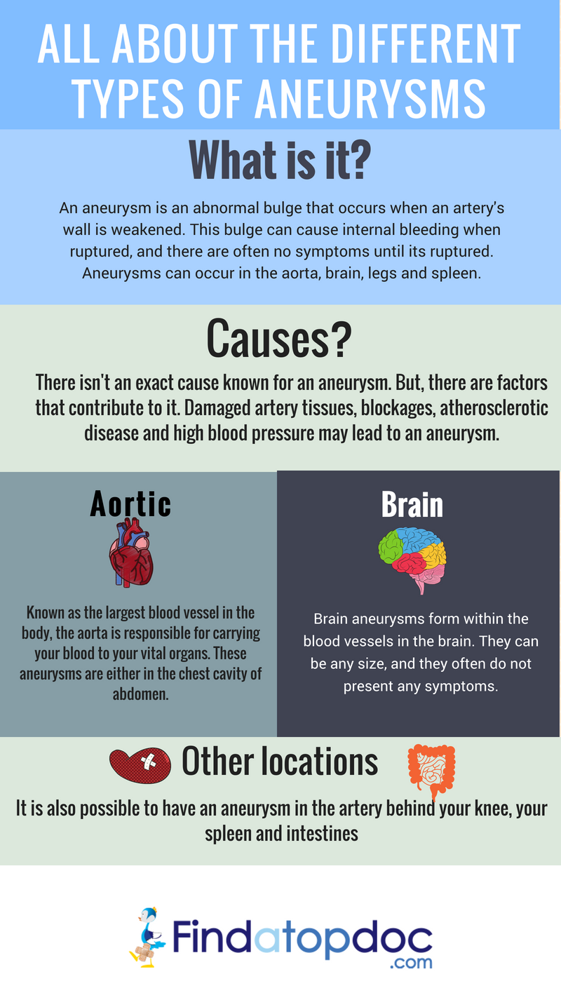
There are different types of aneurysms:
Aortic aneurysm – this aneurysm is formed in aorta, the largest blood vessel in the body. It is categorized into thoracic aneurysm and abdominal aneurysm, based on location.
Brain aneurysm – in this type, the aneurysm is seen in the blood vessel carrying blood to the brain. Rupture of brain aneurysms cause bleeding in the brain.
Peripheral aneurysm – these are seen in other blood vessels like those of the leg, groin, or neck.
Aneurysms may develop slowly over many years. Small aneurysms may remain without any obvious symptoms.
Aneurysms near the skin surface are characterized by throbbing lumps with pain and swelling.
Un-ruptured aneurysms in the brain may sometimes press against the surrounding nervous tissue causing symptoms like blurred or double vision, dizziness, drooping eye lids, and severe headache.
Large abdominal aneurysm cause lower back pain, abdominal pain, or a pulsating sensation in the abdomen. If the aneurysm press on the vagus nerve, it result in coughing, wheezing, and difficulty in swallowing.
Rupture of thoracic aortic aneurysm causes a sharp, severe pain in the chest. The pain may be felt in the jaw, neck, and arms.
Ruptured brain aneurysm causes bleeding in the brain characterized by symptoms like
Deep wounds, injuries, or infections of blood vessels
Genetic abnormalities
Some inherited diseases like Marfan syndrome
4 Making a Diagnosis
Aneurysm is usually diagnosed during routine screening or a checkup for another condition.
X-ray images of the thoracic region taken for some other reasons reveal aortic aneurysms.
Echocardiogram captures the images of the different parts of aorta and the abnormalities, if any.
MRI is another imaging technique that is recommended to visualize abnormalities in the blood vessels. In most of the cases the aneurysm is detected only after the rupture of the bulge.
CT scans help to visualize the blood vessels and bleeding. In CT angiography, a dye is injected to help locate the aneurysm or rupture. When the blood vessel ruptures, red blood cells can be found in the fluid surrounding the brain, called cerebrospinal fluid.
Cerebrospinal fluid test that shows the presence of red blood cells is a positive indicator of rupture within brain.
Cerebral angiogram is another imaging technique commonly used in detecting brain aneurysms. In this procedure, a special dye is injected into the arteries. It travels throughout the brain and helps to make clear images of the blood vessels in brain.
Treatment depend on the location and size of the aneurysm, and overall health of the patient.
Aneurysms in the lower chest and abdominal part of aorta is not life-threatening and is not treated immediately. If they grow rapidly and become large enough to produce symptoms, surgery is suggested.
Surgery is suggested for peripheral aneurysms, where the weak section of the blood vessel is replaced with a graft. For small and stable aneurysms in abdominal aorta and lower chest, regular checkups to monitor the growth of the bulge, is recommended.
Beta blockers are prescribed to keep blood pressure under control. This reduces the pressure on aortic walls and the chance of blood vessel rupture.
Two major treatment methods of ruptured aneurysm are surgical clipping and endovascular coiling.
Surgical clipping
In this procedure, the aneurysm is accessed by opening the skull. A small clip is then placed at the neck of the bulge to prevent blood flow to the aneurysm. This prevents leakage or rupture of aneurysm.
Endovascular coiling
This is a less invasive procedure in which a catheter is inserted into an artery and led to the aneurysm. The catheter is usually inserted into an artery in the groin. A soft platinum wired is pushed into the bulge with the help of the catheter. The soft wire coils in the bulge and blocks the blood flow. This causes the blood to clot that seals the aneurysm completely.
6 Prevention
Lowering the risk factors is the best way to prevent aneurysms and its complications.
Your FindaTopDoc account is completely free. Find doctors & request online appointments. Participate in Health Journeys in over 100 specialty communities. Easy & secure access! Simple Facebook login.
FindATopDoc is a trusted resource for patients to find the top doctors in their area. Be visible and accessible with your up to date contact
information, certified patients reviews and online appointment booking functionality.