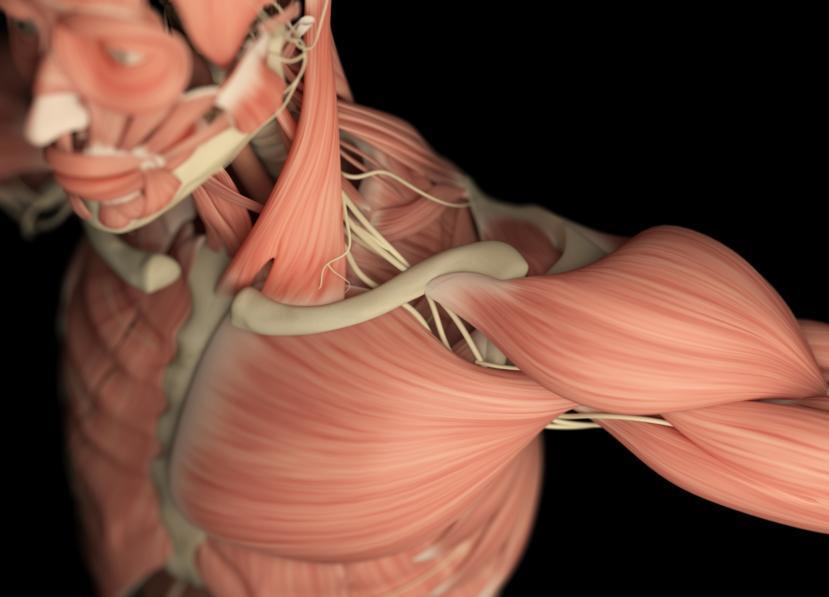
Muscular Dystrophy: A New Understanding of How Muscles Work

A new technique out of McGill may be the key to shed new light on how muscles work. It’s an important step towards a better understanding of how muscle-targeted diseases and disorders work. With a greater understanding also comes a better handle of how to treat and possibly cure these diseases in conjunction with newer technology and genetic tools. The study took a more in depth look at how sarcomeres, the building blocks of the skeletal and cardiac muscles in the human body work together.
Most people never think of how much they use the muscles. Everything that is done requires use of the muscles, from speaking to simply sitting and stretching your legs. The body is made up three different types of muscles, smooth muscle, cardiac muscle and skeletal muscle.
Smooth Muscle
This type of muscle is found in blood vessels, the eyeballs, arteries, the digestive system, and the reproductive organs. Smooth muscle is so called because it can stretch and maintain tension for extended periods of time for example the uterus of a pregnant woman, and it is controlled by the nervous system.
Cardiac Muscle
Cardiac muscle is found only in the heart. It has limited stretching ability like smooth muscle and is what is known as a slow twitch muscle meaning it meant for endurance. It is also able to contract with the force of skeletal muscle, but contracts involuntarily.
Skeletal Muscle
This is the type of muscle that can be felt, think biceps and seen if someone flexes that bicep. When we exercise to build muscle, this is the type of muscle that we build. They’re called skeletal muscles because they attach all over the skeleton. Skeletal muscles are paired for use, one muscle will move bone in one direction, while its pair will move the bone in the opposite direction. Unlike cardiac muscle which is shares contraction force, skeletal muscle can and will contract voluntarily. Skeletal muscle is the type of muscle that is featured in the McGill study, specifically the millions of tiny striated muscles in the body that are known as sarcomeres.
What are Sarcomeres?
A sarcomere is a unit of striated muscle in the human body. Sarcomeres are the basic building blocks for most of the muscles in the body. Some of the muscles of the body is made up of bundles of muscle fibers and these are comprised of smaller strands called microfibrils. Each microfibril is in turn made up of two kinds of filaments, a thick and a thin strand organized in regular, repeating subunits, these subunits are known individually as a sarcomere. The majority of the muscles in the body are of the striated variety making them sarcomeres, this is why when people have degenerative muscle diseases like Parkinson's of types of muscular dystrophy, the result is an overall destruction of the muscles to the point of paralysis.
The sarcomeres capacity for contraction is what makes it important to skeletal muscle features, the thick and thin filaments are what do the work of the a muscle. The protein myosin arranged in a cylindrical shape on the molecular level is what makes up a thick filament. Actin is the protein arranged in the same fashion that make up thin filaments that look like two strands of pearls twisted around each other.
How Sarcomeres Work?
When muscles contract, this is the starting point of what makes work and allow us movement in any way that we would like to. Any action that is undertaken by the body, whether raising your arms or turning your head is a result of muscle contractions. During contractions the thick fibers pull thin filaments past them which makes the sarcomeres shorter (contraction), in the muscle fiber the contraction signal causes contraction all over the so that all the microfibrils shorten simultaneously Inside of each thin filament are two rod like proteins, troponin and tropomyosin that are the molecular switches controlling the interaction of myosin and actin and what causes the filaments to slide along the thick filaments.
But this doesn’t explain how the force needed to make the muscles work is created, just how the process of contraction happens. Force starts with contraction, and these happen when the thick filaments grab hold of the thin filaments and this creates what is known as a crossbridge, muscles then create force by cycling a crossbridge. For example, when contraction begins the myosin molecule forms a chemical bond with an actin molecule creating the crossbridge. In this process a number of other molecules become involved, namely adenosine diphosphate (ADP) and inorganic phosphate (Pi). As soon as the crossbridge is formed the golf club shaped myosin head bends creating force and sliding the thin filament past the myosin in a process called a power stroke. The myosin release the ADP and Pi during the power stroke and a molecule of adenosine triphosphate (ATP) binds to the myosin. When ATP binds, myosin then releases the actin, the ATP molecule splits into ADP and Pi. The energy from ATP resets the myosin head to start all over again. The is the process of cycling a crossbridge that creates the force that muscles need to move and it happens in every sarcomere.
What is Needed for These Processes to Work?
All muscles are triggered by electricity, usually internally by nerve cells in a healthy properly working body or externally with devices such as pacemakers. Muscle contractions are regulated by what the level of calcium ion is in cytoplasm. In skeletal muscle they move a troponin-tropomyosin complex from the binding site allowing actin and myosin to interact. In order to do this effectively, it needs energy. As we said previously that energy is supplied by ATP which is created by the muscles breaking down creatine phosphate and then adding the phosphate to ADP and Pi. Anaerobic respiration then occurs and this is the process by which glucose becomes lactic acid and ATP is made or Aerobic respiration takes place by breaking down glucose, glycogen, amino acids and fats are broken down if oxygen is present to create ATP.
What the Research Into Sarcomere Cooperation Means?
The research out of McGill also points to a cooperation between sarcomeres that was previously unknown. When they examined the microscopic sarcomeres, they discovered that in the healthy myofibril all the sarcomeres in close proximity adjusted to the activation of one sarcomere. The researchers believe this to be crucial to understanding the molecular mechanism of contraction which opens up many new avenues of possibilities in the muscle field. They plan to continue their research to try and discover what happens when sarcomeres fail to cooperate.
Many diseases can affect the balance between contraction and relaxation of muscles. Being the building blocks of the muscle system of skeletal muscle means that sarcomeres are in a unique position to limit those disruptions. The use of modulators can be applied in various ways to correct the imbalances directly or indirectly either through signal pathways or interaction with the proteins that control contractions. That could be a breakthrough in treating such hereditary skeletal muscle disease such as the different forms of muscular dystrophy, heart disease, cardiomyopathies as well as others.


